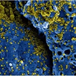
SEM of iron oxide nanoparticles
Sample preparation
Materials
Iron oxide nanoparticles
Ethanol
Deionized water
Carbon-coated copper grids
Ultrasonic bath
Centrifuge
Oven
Protocol
Prepare the nanoparticle suspension by diluting the iron oxide nanoparticles in ethanol to a concentration of 1 mg/mL. Sonicate the solution for 10 minutes to disperse the nanoparticles evenly in the solvent.
- Place a carbon-coated copper grid onto a clean surface, such as a piece of filter paper or a clean petri dish.
- Pipette 50 µL of the nanoparticle suspension onto the grid, making sure to distribute the liquid evenly over the surface of the grid.
- Allow the nanoparticle solution to settle onto the grid for 10 minutes. This will allow the nanoparticles to adhere to the grid and form a uniform layer.
- Gently rinse the grid with deionized water to remove any excess ethanol and let it air-dry for 30 minutes at room temperature. Alternatively, the grid can be dried using a low-pressure stream of nitrogen gas.
- Place the grid into an ultrasonic bath filled with deionized water for 5 minutes to remove any remaining impurities and debris. This will help to ensure that the grid is free of any contaminants that could interfere with SEM imaging.
- Remove the grid from the ultrasonic bath and let it air-dry for 10 minutes at room temperature.
- The grid is now ready for SEM imaging. Place the grid onto the SEM sample holder, making sure it is securely fastened.
- To enhance the contrast of the nanoparticles, the grid can be sputter-coated with a thin layer of metal, such as gold or platinum, using a sputter coater. This will provide a conductive surface that will enhance the signal from the SEM detector.
- Insert the sample holder into the SEM chamber and image the iron oxide nanoparticles using the desired SEM parameters.
- Once imaging is complete, remove the grid from the SEM and store it in a clean, dry container until further use.
Notes
It is important to handle the nanoparticles and grid carefully to avoid any contamination or damage.
The concentration of nanoparticles in the ethanol solution can be adjusted based on the desired density of particles on the grid.
The drying time can be adjusted based on the humidity and temperature of the environment.
The ultrasonic bath helps to remove any debris or impurities that may interfere with the SEM imaging.
The sputter coating step can be omitted if the nanoparticles have a high contrast on the carbon-coated grid.
References
Cullity, B. D., & Graham, C. D. (2009). Introduction to magnetic materials. John Wiley & Sons.
Singh, M., Singh, N., & Singh, R. P. (2018). Iron oxide nanoparticles: Synthesis, functionalization, and biomedical applications. In Nanomaterials in tissue engineering (pp. 31-56). Elsevier.
Tassaing, T., Vayer, M., Bouguelia, S., Gautron, E., Lacroix, L. M., & Bertrand, J. (2017). Synthesis and characterization of iron oxide nanoparticles for biomedical applications: A review. International Journal of Pharmaceutics, 532(1), 101-114.

How to obtain the best SEM images of iron oxide nanoparticles?
Use a low accelerating voltage: Iron oxide nanoparticles are sensitive to high-energy electron beams and can be easily damaged or destroyed by them. Therefore, it is recommended to use a low accelerating voltage (e.g. 5 kV or lower) to minimize damage to the sample.
Optimize the working distance: The working distance is the distance between the sample and the SEM detector. It is important to optimize this distance to obtain the best possible image quality. Too high or too low of a working distance can result in blurry or distorted images.
Use a high-resolution detector: To obtain high-resolution SEM images of iron oxide nanoparticles, it is recommended to use a high-resolution detector, such as a field emission gun (FEG) SEM or a scanning transmission electron microscope (STEM).
Prepare the sample carefully: Proper sample preparation is crucial for obtaining good SEM images. The sample should be evenly distributed on the grid and free of any debris or impurities that could interfere with imaging.
Avoid charging artifacts: Iron oxide nanoparticles can be prone to charging artifacts, which can cause distortion or blurring in the SEM images. To avoid this, the sample can be coated with a thin layer of conductive material, such as gold or platinum.
Experiment with different imaging parameters: SEM imaging parameters such as magnification, contrast, and brightness can be adjusted to optimize the image quality. Experiment with different parameters to find the best settings for your sample.
Take multiple images: To ensure that you capture all of the important features of the sample, take multiple SEM images from different angles and at different magnifications. This will help to ensure that you obtain a complete and accurate picture of the sample.
Interpret the results with caution: Finally, it is important to interpret the SEM images of iron oxide nanoparticles with caution. The images may contain artifacts or other features that could be mistaken for nanoparticles. It is important to use other analytical techniques, such as TEM or X-ray diffraction, to confirm the presence of iron oxide nanoparticles in the sample.

Why Choose Us?
- 24/7 Service
- Accurate Analysis by Expert Scientists
- Free revisions After Completion of Orders
- Guaranteed Satisfaction
- Affordable Price
Advanced SEM Solutions: Imaging, Coloring, and Analysis
We understand that obtaining clear and accurate images of your samples is crucial for your research and development, and we are here to help you achieve your goals.
Our team of experienced professionals has the expertise and skills needed to perform the most advanced SEM imaging and analysis techniques. We use state-of-the-art equipment and cutting-edge software to provide you with the best possible images of your samples.
We can help you with the following services:
Image enhancement: We can enhance the quality of your SEM images by adjusting the contrast, brightness, and other imaging parameters to make the features of your samples more visible.
Coloring: We can color your SEM images to highlight specific features of your samples, making them easier to interpret and understand.
Analysis: We can analyze your SEM images using advanced software tools to identify and quantify the features of your samples, such as particle size and distribution.
Report generation: We can generate detailed reports of our analysis results, including statistical data and visual representations of the features of your samples.

Any Questions?
We are confident that our services will help you to achieve your research and development goals by providing you with high-quality SEM images that are accurate and easy to interpret. By partnering with us, you can save time and resources, allowing you to focus on other important aspects of your research.
In addition, we offer competitive pricing and fast turnaround times, ensuring that you receive your results quickly and at an affordable price. We also provide excellent customer service, working closely with you to ensure that your needs are met and your expectations are exceeded.
We believe that our services will be of great value to your organization, and we look forward to the opportunity to work with you. Please contact us today to learn more about how we can help with your SEM imaging and analysis needs.