Colored SEM

colored SEM Bacteria

Are SEM images colored?

Because it is one of the most versatile imaging and measurement tools, the scanning electron microscope (SEM) is widely used in many fields of industry and science. Images produced are praised for their high depth of field and excellent image resolution, which are orders of magnitude better than light microscopy. However, the traditional SEM only provides black and white images, and each image only contains information in two dimensions. Since SEM creates images using electrons rather than visible light photons, grayscale images are obviously common.

Researchers colorize SEM images of the nanoscopic world in order to more effectively explain their findings. Colored SEM images are frequently used in research papers. According to reports published in Science magazine, researchers can currently produce colored SEMs while imaging. Every time a metal loses an electron, the microscope notices it and records it as a distinct loss of color. Colored SEM images are very demanding and Artificial coloring systems are expected to be included in future generations of electron microscopes.

SEMs that have been artificially colored should not be confused with the Energy dispersive X-ray spectroscopy (EDX) technique. Energy dispersive X-ray spectroscopy is a technique for elemental analysis and determining chemical composition that is related to electron microscopy. The method is based on the production of characteristic X-rays, which reveal the identity of the elements present in the sample. Typically, researchers use this technique to color SEM images based on the types of elements in the material.

Why should I colorize SEM images?

SEM images are widely used in different fields of study, ranging from chemistry to biology. SEM images are black and white by nature and adding multiple colors to them is a great idea for easier object identification and differentiation. Colored SEM images are highly recommended to help distinguish the different things in the electron microscopy image.
Many prestigious journals recommend colorizing SEM images for enhancing visual illustration. False-colored SEM images make your research more eye-catching. The colored SEMs look more realistic and color can help the readers of your publication distinguish the different features in the image. Many prestigious journals feature works with colorized SEM image on their front cover

What are examples of colored SEM?

colored SEM - COVID19
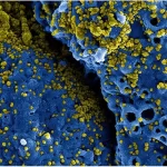
Colorized electron microscopy images can be used in various fields including biology, medicine, nanotechnology, and material science. For example, colored electron microscopy images have been used in articles to illustrate the structure and function of coronaviruses. 

How to add color to SEM images?

To colorize SEM images, some researchers use specialized software created for this purpose, while others use professional design software like Photoshop. The best option is to have the SEM images colored by experts so that, rather than wasting hours of your valuable time and money on pricey software, you can get a professionally colorized SEM image quickly and at a reasonable cost.

In Unitechlink Inc., Our expert scientists work with our art departments to color your SEM and TEM images and make them incredibly charming! Also, we are able to convert standard SEM images into 3D-like images (that is, those representing X, Y, and Z (height) coordinates).

Limited Offer: Colorize Your SEM Image for Just $35 with Unlimited Revisions Until Satisfaction!

Why Choose Us?

Please fill out the form below, and we will contact you shortly!