

SEM Colouring Gallery
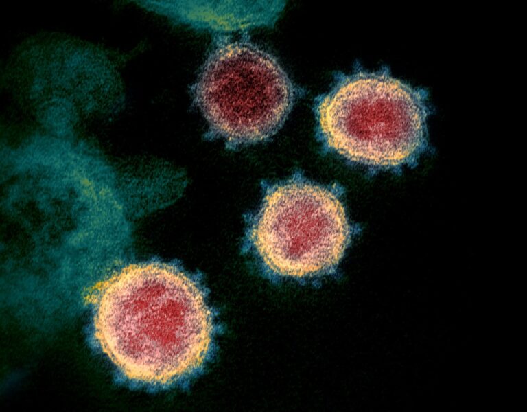
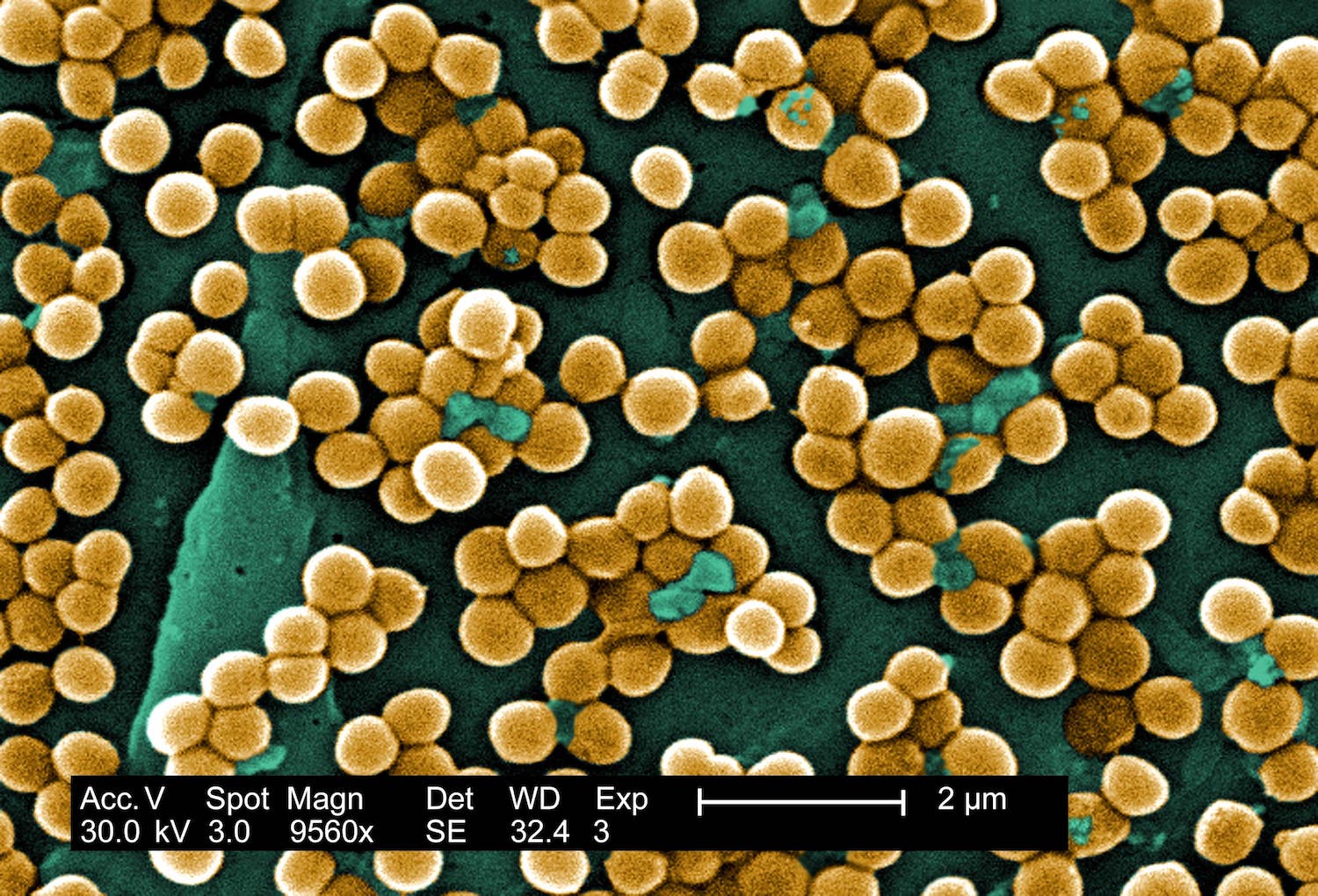
Examples of colored electron microscopy visuals that show how strategic image enhancement can help reveal structures, add visual depth, and improve figure presentation.

High-impact SEM visualization for improved structural clarity

Scientific coloring for stronger visual separation
Publication-ready microscopy figures with expert finishing
Ideal for journals, lab websites, posters, and presentations