Professional SEM image enhancement services
Professional SEM-focused image enhancement and colorization services, with TEM visualization available. Enhance clarity, highlight structures, and prepare publication-ready electron microscopy images with expert precision.


What this service helps you achieve
Our SEM-focused electron microscopy image enhancement workflow also supports TEM visualization. It is designed for scientific communication, stronger visual impact, and clearer structure interpretation without making your figures look overprocessed or artificial.
Enhance clarity
Improve figure readability by separating features, reducing visual ambiguity, and creating a cleaner image hierarchy for presentations, manuscripts, websites, and grant applications.
Highlight structures
Use careful scientific coloring and contrast guidance to emphasize morphology, particles, microbes, interfaces, or regions of interest so key scientific details are easier to communicate.
Prepare publication-ready visuals
Create polished microscopy figures suitable for research groups, journal submissions, posters, slide decks, scientific outreach, and educational materials.
Why professional SEM coloring matters
Professional colorization helps transform black-and-white microscopy images into clearer, more engaging, and easier-to-understand scientific visuals.
SEM images are widely used across different fields of study, ranging from chemistry to biology. SEM images are black and white by nature, and adding multiple colors can be a great way to support easier object identification and differentiation. Colored SEM images help distinguish different structures within electron microscopy images and can enhance visual illustration for scientific communication. False-colored SEM images can make research figures more eye-catching, while carefully selected colors help readers distinguish important features in the image. Many prestigious journals and scientific publications feature colorized SEM images because they can improve clarity, visual impact, and overall presentation quality.
Professional SEM/TEM colorization
- Below 3 images: $50 USD per SEM/TEM image
- Above 3 images: $30 USD per SEM/TEM image
- Offer: 2 free coloring services for more than 10 SEM images
- Designed for stronger clarity and visual communication
- Ideal for publications, posters, talks, websites, and lab profiles
- Email-based ordering or direct order through the website
How to order
By email: service@unitechlink.com
Online: Use the order form below
Send your SEM or TEM image, describe the areas you want emphasized, and mention where the image will be used, such as a journal article, thesis, poster, website, or grant deck.
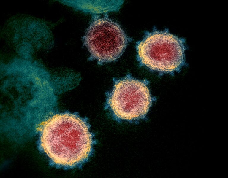
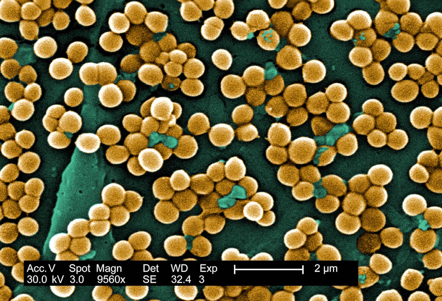
Start Your OrderSample SEM image enhancement gallery
Examples of colored electron microscopy visuals that show how strategic image enhancement can help reveal structures, add visual depth, and improve figure presentation.
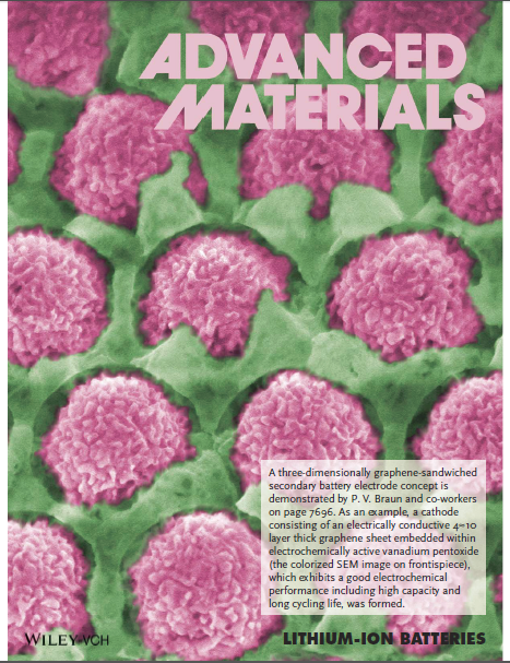
Journal-cover visual inspiration
Microscopy images can become high-impact scientific visuals when structure, contrast, and composition are handled carefully. The examples below are included as journal-cover style references only.

Built for leading research environments
This service is designed for the needs of faculty members, postdoctoral researchers, graduate students, core facilities, microscopy labs, biotech teams, and scientific communicators who need polished SEM and TEM visuals with stronger presentation value.
Simple 4-step workflow
A fast and low-friction process for labs, faculty members, students, and research teams that need polished microscopy visuals without wasting time.
Send us a message
Email us at service@unitechlink.com or submit through the order form below to start your SEM/TEM image enhancement request.
Share your priorities
Tell us which features matter most, such as particles, cells, bacteria, surfaces, or background separation.
Get your image
Receive your enhanced SEM/TEM image prepared for clearer scientific communication and stronger presentation value.
Receive unlimited revisions
Receive unlimited revisions until satisfaction, so the final image matches your scientific and presentation needs.
What researchers say
Feedback from real use-cases in academic and research environments.
Serving researchers across leading world universities
This service is suitable for researchers and labs working in top academic environments around the world.
Frequently asked questions
Helpful answers for researchers, labs, and students considering SEM or TEM image enhancement services.
What types of images can I send?
You can send SEM or TEM images intended for research papers, conference posters, presentations, lab websites, grant materials, teaching, or outreach.
How much does it cost?
Below 3 images: $50 USD per SEM/TEM image. Above 3 images: $30 USD per SEM/TEM image. Offer: receive 2 free coloring services for orders of more than 10 SEM images.
How do I place an order?
You can order by email at service@unitechlink.com or through the order form at the bottom of this page.
Why use SEM/TEM image enhancement?
Strategic coloring and contrast enhancement can improve readability, distinguish structures, add visual hierarchy, and make microscopy figures more compelling for scientific communication.
Can this help with publications?
Yes. The service is specifically presented as useful for publication-ready visuals, conference figures, posters, presentations, and scientific outreach assets.
Ready to enhance your SEM or TEM image?
Order by email or submit directly through the form below. Clear pricing, academic-friendly communication, and polished SEM-focused electron microscopy visuals with TEM support.
Order form
Please complete the form below to submit your SEM/TEM image enhancement request. You can also email us directly at service@unitechlink.com.